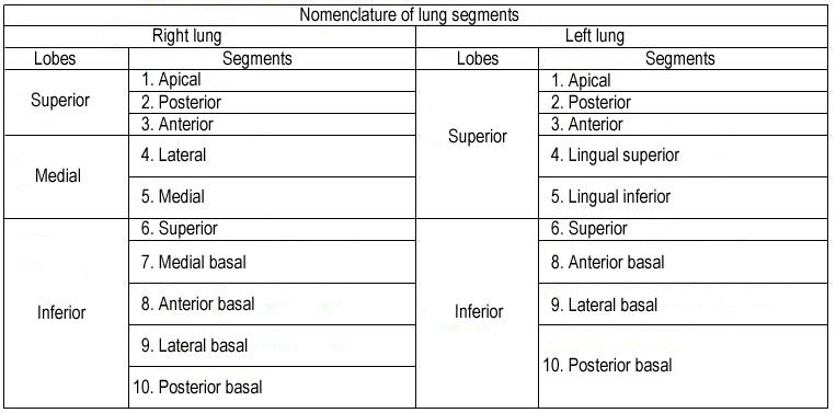
The lungs comprise from the lobes. The right lung has 3 lobes, (superior, medial, inferior), left – 2 lobes (superior and inferior). The lobes divide into the segments. In the right lung there are 10 segments and in left lung – 9 (see tab. 1.4.1.). The segments comprise from lobules. In both lungs, there are about 1000 lobules. In general the size of lobule is 1 – 1.5 cm. The collection of the lobules comprise subsegment. Several subsegments comprise a segment.
The lung’s segment topography.
Each lung’s segment has bronchus and artery, and both bronchi and arteries are arranged almost parallel. The arrangement of the vein segments are not identical with bronchi-arterial, though under the form resemble them. The bronchi-lung segments under the form remind a triangle, top directed medial, and basis – to peripheral surface. Each lung segment is separated from next by a layer of connecting tissue.
Bronchial Tubes.
The two bronchi proceed from the bifurcation of the trachea opposite the 4-th dorsal vertebra to their correspond¬ing lungs. Upon entering the lungs, the bronchus divide into branches and each of these divides and subdivides dichotomously to their ultimate termination (smallest bronchia).
The structure of the lung parenchyma.
The finniest, independent functional unit of the lung parenchyma is an acinus. It is a miniature lung in diameter about 1,5 mm. The acinus is ventilated by the smallest bronchioles (bronchiolus or bronchulus terminalis) – finniest branching of the bronchial tree. The group of acinus forms lobulus, which diameter reaches1 – 1.5 cm (fig. 1.4.1.).
The mucous membrane lining the bronchial tubes, provided with a ciliated columnar epithelium as far as their termination; but in the alveolar passages and air-cells it is altered in its characters, is thin and transparent and coated with a squamous epithelium.
The lung blood vessels.
The pulmonary artery, conveying the dark and impure venous blood to the lungs, accompanies the bronchial tubes to the lung, and divides as the tubes divides. The branches terminate in capillary vessels, which-form a dense network in the parietals of the alveolar passages and air-cells, and then converge to form the pulmonary veins, by which the arterial blood, purified in its passage through the capillaries, is returned to the left auricle of the heart. In their course through the lung, the artery is commonly found above and behind the bronchial tube, while the vein is below and in front.
Pleura.
Each lung is enclosed and its structure supported by a serous membrane, the pleura, which invests it as far as the root, and is then reflected on the parietes of the chest. That portion of the membrane which is in relation with the lung is called (pleura visceralis s. pleura pulmonalis), and that in contact with the parietes, pleura costalis, pleura diaphragmatica and (pleura mediastinalis).. The pulmonary pleura is very thin, is elastic, and inseparably connected with the structure of the lung; the costal pleura is thick and strong, has very little elasticity, and can be readily stripped off the ribs and intercostal muscles which it covers.
The lymphatic lung system.
The lymphatics on the surface of the lung form a fine sub-pleural plexus, communicating by means of stomata with the pleural cavity. Those in the substance of the lung consist of two sets, one of which forms an elaborate plexus beneath the mucous membrane of the bronchial tubes, and the other originates in capillaries between the air-cells and alveolar passages. They all terminate in the bronchial glands of the root of the lung. These glands, very numerous and often of large size, are placed at the roots of the lungs, around the bronchi, and at the bifurcation of the trachea.
The lung nerves are derived from the pneumogastric (parasympathetic) and sympathetic nerve system. They form two plexuses: anterior pulmonary plexus, situated upon the front of the root of the lungs, and composed chiefly of fila-ments from the deep cardiac plexus; and posterior pulmonary plexus, on the posterior aspect of the root of the lungs, composed principally of branches from the pneumogastric (n.vagus). The branches from these plexuses follow the course of the bronchial tubes, and are distributed to the intercellular passages and air-cells.
Mediastinum.
The two pleural sacs do not communicate with each other, but have between them a space which contains all the viscera of the chest with the exception of the lungs. This is called the inter-pleural space or mediastinum. The mediastinum is divided into an anterior, middle, posterior, and superior portions.
Lung function.
The functions of lungs are closely connected to features of their structure. Due to the presence of hundreds and millions of alveoli, total surface of which reaches 100 m2, the networks of capillaries with a surface up to 80 m2, together with components of alveolar -capillary membrane. The lungs serve not only for breath, but also for expectoration, for maintenance of a constancy of the body temperature. The lungs participate in development of substances participating in regulation of blood clotting, in metabolism of proteins, fats, and carbohydrates.