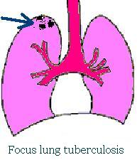
Focus lung tuberculosis makes about 50 % of all newly reveled tuberculosis forms. The course of this form can proceed without subjective sensations and occasionally it is detected by mass fluorography. After subsequent medical examination it is quite often reveals, that patients did not pay attention to symptoms of tubercular intoxication for rather long time.
According to clinical and radiographic picture two forms of focus tuberculosis are distinguished: soft (acute) focus tuberculosis and fibrous focus tuberculosis. In the process of healing of the different forms of tuberculosis residual focuses are formed. These focuses are replaced by fibrotic tissue, being encapsulated and are considered as fibrotic residual focuses. The pathogenesis of focus tuberculosis is various, uncomplicated and complicated. Focus tuberculosis can be derived action of the primary or secondary period of tuberculosis. The focal forms of secondary tuberculosis arise among adults under influence of super exogenous infection or endogenic dissemination of MBT, from previously latent focuses. Such foci contain caseous material and are located in lymphatic nodes, scars, places of indurations in the lungs or in other organs.
In period of acute condition of the disease MBT disseminate from focuses along lymphatic pathways and fine bronchi. More often fresh focuses occur in upper lobes of the lungs. At the beginning, in the places where MBT settled endobronchitis is developed. Then the inflammation covers all fine branches of bronchi. Caseous necrosis develops in damaged bronchi with the subsequent transition into pulmonary tissue, more often to apical areas. The small focus develops as caseous, acinus or lobular pneumonia. Lymphatic network is involved into pathological process only around of the focus. Regional lymphatic nodes do not react on inflammation in the lungs. Exudations are insignificant and are quickly replaced by productive reaction.
Hematogenic dissemination is characteristic by symmetric localization of the focuses, which rests in apical areas of the lungs.
Clinical picture.
 The part of the patients revealed by fluorography really has no any clinical symptoms. However majority of the patients react to occurrence of focus lung tuberculosis with weakness, sweating, downturn of work capacity and appetite. Patients complain of occurrence heat in cheeks and palms, on short-term shivering and small subfebrile temperature during a day. The unstable cough — dry or with poor amount of sputum, pains in the sides are observed sometimes. At examination of the patient weak pain is marked of muscles humeral zone on the side of inflammation. Lymphatic nodes usually do not react. In lungs can be revealed shortening of percussion sounds only when focuses of inflammation begin to merge. In acute phases of focus tuberculosis development and at presence of infiltrations at coughing out are listened harsh breath and rare fine damp rattles. Tuberculin tests are usually moderately expressed.
Changes in the blood anything characteristic for this form of disease are not marked, and the changes of blood depend on a phase of disease. At less severe forms blood is normal. In a phase of infiltration ESR is a little bit accelerated, the left shift of the stub neutrophile formula reaches 12 — 15 %, insignificant lymphopenia.
At sub acute development of the process the so-called productive form of focus tuberculosis is observed. The focuses are in average (3 – 6 mm), round or, irregular form precisely outlined, of average or sharp intensity is defined. The focuses are localized separately, do not merge. Around focuses it is possible to visualize lymphangitis as thin rods and net like picture.
Treatment.
At modern antibacterial treatment the fresh tubercular focuses and resolution of lymphangitis usually lasts for 12 months, more often with complete restoration of pulmonary structure or remains insignificant pronounced pulmonary structure and fine outlined focuses. Less often after effective treatment the fresh focuses are not resolved but are becoming encapsulated. At effective treatment much more often outcome of inflammation ends with formation of a capsule, and rough fibrous develops in a place of lymphangitis.